Activation programmed cell death. Sensitization of defense responses and activation of programmed cell death by a pathogen 2019-03-25
Programmed cell death

The stimuli can originate intracellularly from within the cell or extracellularly. The relative lengths of various domains and subunits are not drawn to size and only serve as a guide here to demonstrate the various types of apoptotic caspases found in C. Annals of the New York Academy of Sciences. Sodium butyrate NaBt , a naturally occurring short chain fatty acid derived from carbohydrate metabolism in the gut, is known to exhibit strong anti-cancer potentials in various human cancer cells; however, its action mechanism is poorly understood. Zingerone; a phenolic alkanone, one of the active components of ginger, possesses multiple biological activities, such as antioxidant and antiinflammatory properties.
Infographic: How Cells Cheat Death
Programmed cell death in animal development and disease. However, cytochrome c is not required for the apoptosome assembly and fails to bind the complex. It is a crucial host defence mechanism, and pathogens have evolved to both evade and co-opt apoptosis. The balance between proliferative and non-proliferative behaviour is very carefully regulated, there being genes involved in growth arrest as well as the better characterised genes involved in mitogenesis. Disruption of mitochondrial membrane integrity and cytochrome c release. For example, while we know that the transcriptional activation of the gene specifies whether a cell will live or die, very little is known about what regulates expression.
Role of NF
Significant reduction of tumor cells proliferation, augmentation of tumor cells apoptosis and suppression of angiogenesis were observed in the combination group compared with controls. This not only avoids the damaging consequences of cell necrosis but also allows the organic components of the dead cell to be recycled by the cell that ingests it. These results indicate that cloned proteasome subunits having similar sequences to the yeast Y13 subunit are structural, but not catalytic, components of proteasome. Following this second centrifugation, the supernatants obtained were taken to represent the cytosol fraction, and the pellets were resuspended in grinding buffer to represent the mitochondria fraction. Apoptosis occurs through the activation of a cell-intrinsic suicide program. Some cells can be killed by engaging certain receptors such as during glucocorticoid-mediated killing of thymocytes. Cell survival and trypan blue exclusion studies indicated that, at low drug concentrations, cells which had transiently arrested in the G2 phase survived, while at higher concentrations only a limited number of survivors were responsible for the observed recovery of growth.
Programmed Cell Death (Apoptosis)
Found to be highly effective against multiple myeloma, bortezomib will soon be tested with first-line anticancer drugs against a broad range of human malignancies, including solid tumors. First, a carefully regulated increase in cell numbers coupled with, second, the differentiation of the appropriate cell types which are, thirdly, arranged in the appropriate spatial organisation. For example, in normal tissues of metazoan organisms the number of cells with any particular phenotype is very carefully controlled Hall and Watt 1989a; Hall 1989. The sensitivity of malignant cells to apoptosis, provoked by this synthetic derivative in vitro, deserves further studies in suitable in vivo models. These observations demonstrate that multiple mechanisms of resistance exist, and often several may occur in the same cell line. Both c-myc and c-myb are normally expressed in proliferating myeloblasts and suppressed following induction of differentiation. Last revised April 11, 2005.
Activation of the programmed cell death pathway by inhibition of proteasome function in plants — Korea University
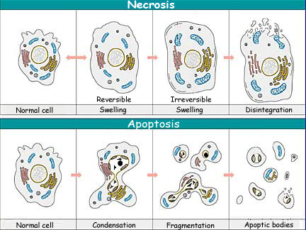
Plasma membrane blebbing and disintegration of the cell into numerous apoptotic bodies are other highly characteristic features of apoptosis Figure 3. Relatively little attention has been paid to the role of cell death in cancer development, probably because for a long time in toxicology cell death was regarded as a passive degenerative phenomenon of secondary importance for the regulation of cell number in tissues. Tens of thousands of new drugs have been screened against this panel and multiple relationships linking growth inhibition, resistance mechanisms, and other genetic characteristics have been elucidated 5,6. The stained leaves were observed under a fluorescence microscope Zeiss Axioskop. This combination promoted a more robust inflammatory response to the tumor that reduced the size of the cancer. Inflammasomes: mechanism of assembly, regulation and signalling. If part of the liver is removed in an adult rat, for example, liver cell proliferation increases to make up the loss.
Programmed cell death protein 1

Rapid induction of inflammatory lipid mediators by the inflammasome in vivo. During apoptosis in animal cells, the release of cytochrome c occurs before visible morphological changes. In this article, I focus primarily on caspases that either have a clearly defined function in apoptosis or have an implied function in cell death pathways. High activity inhibits activity, resulting in the activation of and and the commitment of a cell to the cell death fate. Activation or re-introduction of p53 induces apoptosis in many tumour cells and may provide effective cancer therapy2. We see in the next how these signal molecules help multicellular organisms regulate their cell numbers. The activity of this compound, 1j, was then compared to a popular anticancer drug, cisplatin, having limited usage because of its nephrotoxic nature.
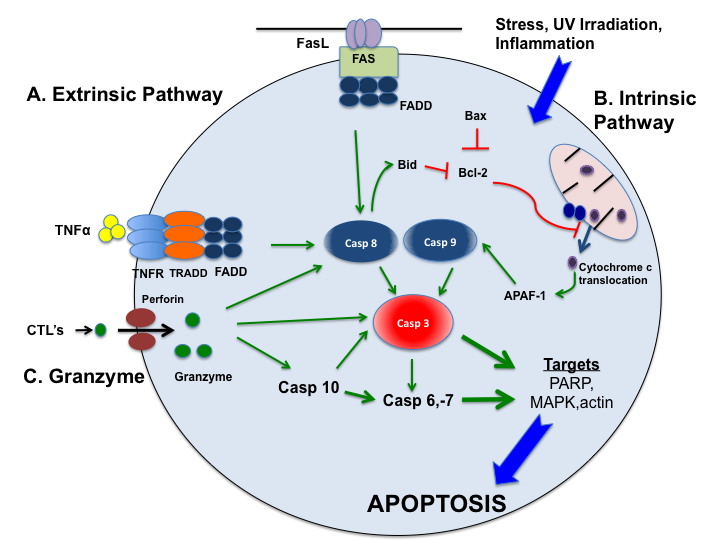
APOPTOSIS(PROGRAMMED CELL DEATH) EXTRINSIC PATHWAY PART 1
Thus, during the past decades increasing evidence has accumulated showing that disturbance of apoptosis is involved in the pathogenesis of tumors Bursch et al. Cell Host Microbe 16, 237—248 2014. Tumor weights were evaluated throughout the treatment duration. Consequently, the loss of genes required for cell death or the over-activation of genes that block it can render tumor cells relatively more resistant to the cytotoxic effects of a broad spectrum of anti-cancer drugs. Given that apoptosis is a fundamental biological process essential for the systematic dismantling and elimination of unwanted cells it is not surprising that impaired apoptosis has been implicated in an increasing number of diseases. The co-polymer layers and their interaction with cancer cells were analyzed by scanning electron microscopy. Even after commencing this suicidal process, cells can recover through a recently discovered process dubbed anastasis.
Caspase function in programmed cell death

In this chapter, we have highlighted many of the basic pathways through which apoptosis can be executed and modulated. Tumor growth was evaluated for 25 days. In the developing vertebrate nervous system, for example, up to half or more of the nerve cells normally die soon after they are formed. Ectopic expression induces translocation from mitochondria to nuclei in a dependent manner, suggesting that the role of mitochondria in regulating apoptosis is conserved. This step would have been risky for the primitive eukaryotic cells, which began to engulf the bacteria, as well as a perilous step for the ancestors of mitochondria, which began to invade their proto-eukaryotic.
(PDF) Activation of the Programmed Cell Death Pathway by Inhibition of Proteasome Function in Plants

For example, is the terminal, global regulator of somatic sexual fate required for female development ; and is essential for viability. Given that there is much in common in the apoptotic function of caspases from various model organisms, I have discussed here work from C. This process of cell death has been identified in the germinal areas of the , , , , and among other regions. Biochemical model for the activation of programmed cell death. It is proposed that the G2 arrest results from the inability of the cells to transcribe genes required for passage into mitosis. However, much remains to be learnt about programmed cell death. Flies mutated at the dredd locus fail to induce the synthesis of antimicrobial peptides and are highly susceptible to infection by Gram-negative bacteria.
Cell
This is an open-access article distributed under the terms of the Creative Commons Attribution License, which permits unrestricted use, distribution, and reproduction in any medium, provided the original author and source are credited. These results mirrored corresponding effects of gene silencing on proteasome activity against peptide substrates. In 1978, Dougherty and coworkers observed that upon injection of hematoporphyrin derivative HpD , a chemically synthesized porphyrin molecule, into cancerous tissues the compound accumulated to higher concentrations in malignant tissue than in normal tissues. This concept eventually helped to elucidate the role of cell death in a variety of patho physiological states Bursch et al. Genetic pathway of programmed cell death in C. Agrobacterium culture for infiltration was prepared as described.