Discography procedure. Discography 2019-05-09
Discography Disc Stimulation Dallas, Addison Texas

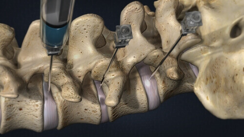
Sterile technique is utilized to minimize the risk of infection. Outside links: For the convenience of our users, RadiologyInfo. Different parts of the body absorb the x-rays in varying degrees. You will likely be instructed not to eat or drink anything after midnight before your procedure. Fluoroscopy, which converts x-rays into video images, is used to watch and guide progress of the procedure. Once inside the disc space, a safe fluid know as contrast dye is injected into the disk.
Discogram (Discography)
The procedure may cause soreness for a few days. If there is any new loss of bladder control or bowel control excessive and uncontrollable amounts , new numbness or weakness, or fever of more than 101 with severe worsening back pain, contact our office immediately, or if it is after hours go to an Emergency Department or Urgent Care, and explain the procedure you have had and the symptoms. Most patients will experience a mild to moderate increase in back pain after discography. Discs degenerate in all adults as we grow older. It is a test to help your surgeon or pain physician to choose options for further treatment.
Discography Video: Non

Psychological factors may be taken into consideration as well. If concordant pain is found in a particular disc, a spine surgeon will then perform a discectomy and fusion. This can leak after injury, and the contents can get into the outer layers or even the nerve roots that are leaving from the lower part of the spinal cord cauda equina. These chemicals may also make the nerve endings more sensitive, so movement that was not painful in a normal disc may cause pain in an injured disc. Modern x-ray systems have very controlled x-ray beams and dose control methods to minimize stray scatter radiation. No randomized, controlled, and blinded studies of discography have been reported. A smaller needle is then inserted through the guide needle and into the center of the disc.
What is Discography?

Most patients who require a discogram have failed to improve with conservative treatments, including medications, physical therapy, and injection therapy. Following discharge home, you should plan on simple rest and relaxation. See the for more information about radiation dose. Low-pressure positive discography in subjects asymptomatic of significant low back pain illness. Others argue that abnormal disc morphology should serve as a gold standard to judge the accuracy of lumbar discography rather than the response to an as yet unproven treatment for a disease difficult to treat. Once the needle is inside the disc, a contrast material is injected and the needle is removed. They usually have had back pain for at least 4 to 6 months.
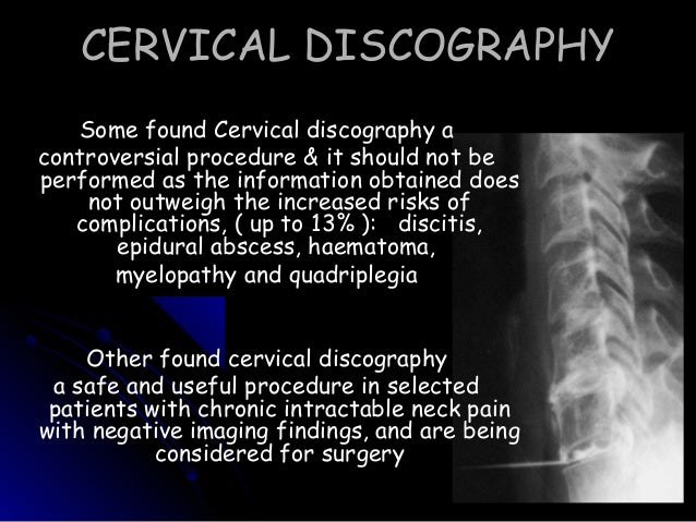
Discography or Discogram to Diagnose Neck or Back Pain
This website does not provide cost information. In some cases, antibiotics are given intravenously before and after the procedure. These stored images are easily accessible for diagnosis and disease management. Pillows may be used to help keep you comfortable and in position. Until recently, x-ray images were maintained on large film sheets much like a large photographic negative.
What is Discography?

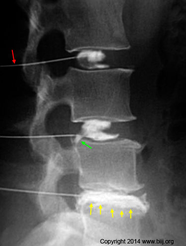
Inserting the Guide Needles For each disc the doctor wants to test, he uses a local anesthetic to numb the skin above your spine and all the tissue down to the disc area. Doctors usually don't rely on the results of a discogram alone because a disk with wear-and-tear change might not cause pain. This gives your health care provider more information about the exact pattern of the spread of the contrast through or out of the disc. These events in the disc may also trigger disc degeneration. X-rays pass through most objects, including the body.
Discography Video: Non
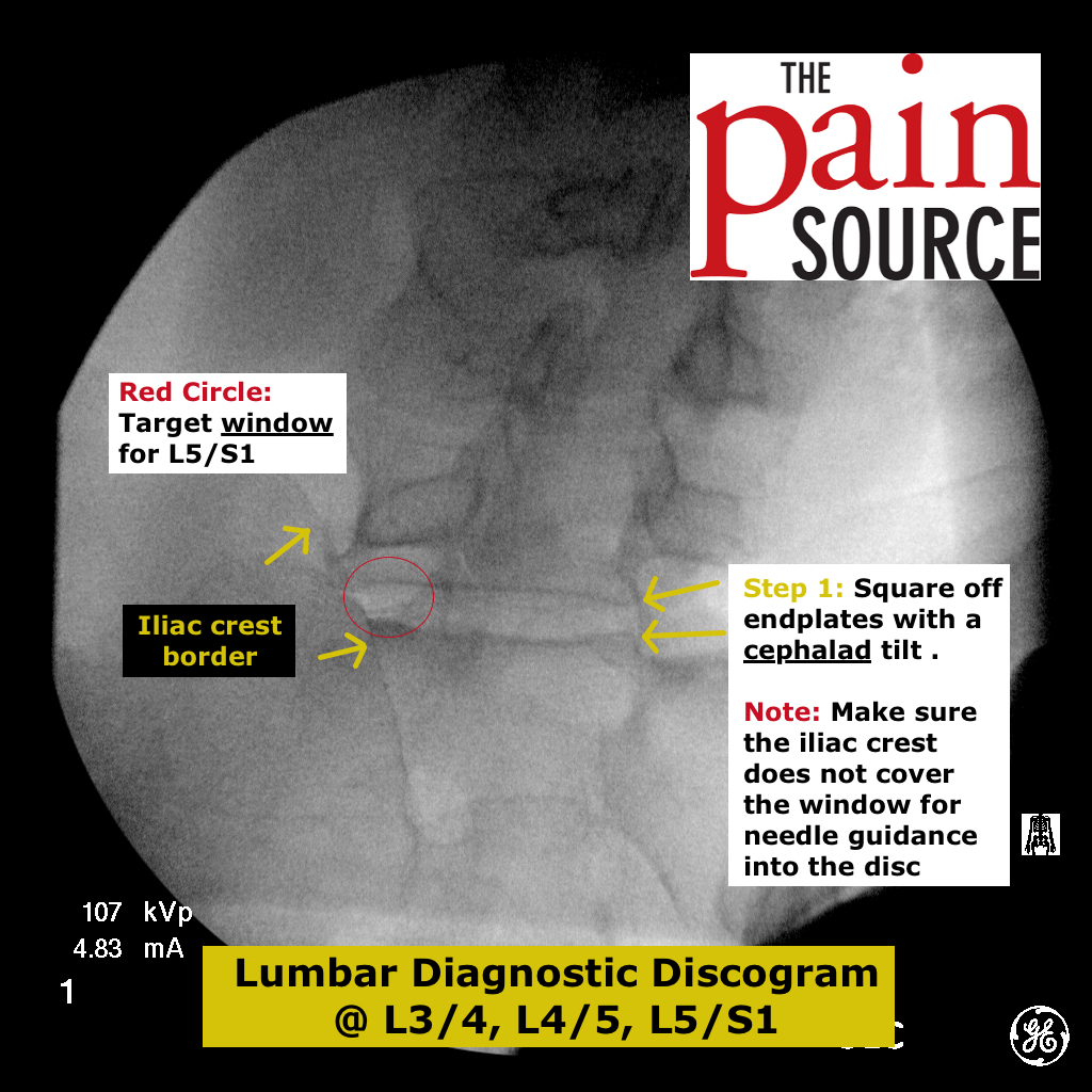
The discography procedure is meant to determine if the abnormalities are causing the pain. After each disc is tested, images are taken with the fluoroscopic unit. With each injection, you will feel either pressure or pain, as shown in illustration 3. If so, the results are used to determine the best type of surgery for you, and which disc levels may be included in surgery. There are a number of factors that indicate you should not have a discogram. Contemporary concepts in spine care: lumbar discography.
Discography Procedure
The pain provoked by the injection should be temporary. Try to rest as much as possible the first several days after the procedure. If you have not already provided your radiologist with prior relevant imaging exams e. Updated review date, Rationale, References and History sections. Removing the Needles After your doctor tests each disc, he uses the fluoroscope to take images of your discs.
Discography at the Southeastern Spine Institute
You should discuss the risks associated with discography and warning signs of complications with your health care provider before the procedure. Because a disc can be damaged without causing pain, the results of a discogram are usually combined with other test results to determine a treatment plan. It also is used to help guide the treatment of abnormal intervertebral discs — sponge-like cushions located between the vertebrae of the spine. In the review of 69 studies meeting a minimal evidence threshold selected by Buenaventura 2007 outlined above, while 39 studies were prospective, only 8 studies were controlled, just 2 were randomized Manchikanti, 2001 and only 1 small observational study Carragee, 2000 used a prospective, controlled, and blinded design. The discography results are reviewed and given to your health care provider.
What is Discography?
Needles are inserted through the back into the near the suspect area, guided by imaging. The authors concluded in their review of the evidence through November 2006 that there is strong evidence for the diagnostic accuracy of discography as an imaging tool, for its ability to evoke pain, and to support the role of discography in identifying a subset of individuals with lumbar discogenic pain. In some cases, or scanning is a better alternative to a discogram for the diagnosis of back pain. The contents of the nucleus of an injured disc may leak out into the outer layers or all the way through those layers to the nearby nerve roots exiting from the cauda equina the lower end of the spinal cord. Because discography is an invasive procedure it involves putting needles into the disc , it is not performed early in the diagnostic and treatment process. The process may be repeated for additional discs. Revised guideline to address lumbar discography only.